
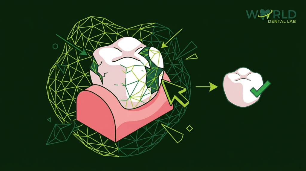
The switch to digital workflows has reduced human error in dental lab production — but it has introduced a new category of error that didn’t exist with physical impressions: STL file problems that are invisible to the naked eye but cause restorations to fit poorly, require remakes, or fail prematurely.
The good news is that most STL file errors are identifiable and fixable before you send the case. This guide covers the 10 most common problems, what causes them, how to spot them, and what to do about each one.
1. Incomplete Margin Capture
What it is: A gap or missing section in the scan data at the preparation margin — the most critical area for fit.
Why it happens: Residual moisture, blood, saliva, or tissue over the margin during scanning. Inadequate retraction. Operator moving too quickly over the margin area.
How to spot it: In your scanner’s preview, rotate the scan and view the preparation from a steep occlusal-to-gingival angle. Any gap in the mesh at the margin level is incomplete capture. Do not proceed if the margin isn’t fully closed in the scan.
Fix: Rescan the preparation. Ensure complete hemostasis and tissue displacement before scanning. Dry the preparation just before scanning each section. Do not attempt to scan a still-bleeding margin.
2. Scan Stitching Errors (Mesh Discontinuities)
What it is: When the scanner fails to align two scan passes correctly, producing a “step” or discontinuity in the mesh — like two puzzle pieces that don’t quite meet.
Why it happens: Moving the scanner too quickly between areas, poor overlap between scan passes, patient movement, reflective surfaces (saliva, metal restorations) disrupting the scanner’s triangulation.
How to spot it: Rotate the scan in all directions and look for steps or seams in what should be a smooth surface. The arch scan should look like a continuous, smooth model — not two halves with a visible join line.
Fix: Delete the affected scan pass and re-scan that section. Slow down through transition areas. Increase drying. On highly reflective metal restorations, use a scan spray to reduce reflectivity.
3. Missing or Incomplete Opposing Arch
What it is: The opposing arch scan is missing, too short, or excludes teeth beyond the preparation area.
Why it happens: Some clinicians scan only the teeth immediately adjacent to the restoration and skip the rest of the arch, assuming the lab only needs local context.
How to spot it: Open the opposing arch scan and check that it covers the full arch (or at minimum the full quadrant), not just 2–3 teeth adjacent to the preparation.
Fix: Rescan the opposing arch in full. The extra 60 seconds at chairside prevents the lab from making occlusal guesses that cost a 30-minute adjustment appointment.
4. Poor Bite Registration Scan
What it is: The bite scan — taken with both arches in occlusion — is incomplete, shows only 2 teeth, or is misaligned.
Why it happens: Rushing through the bite registration step. Not scanning far enough posteriorly. Patient opening slightly during the scan.
How to spot it: In the bite scan, you should see at least 3 pairs of occluding teeth, and the scan should align the upper and lower arches in correct centric relation. If the scan shows only 1–2 tooth pairs, it’s insufficient.
Fix: Rescan the bite, scanning from the buccal corridor across at least the entire quadrant. Instruct the patient to close firmly in their natural bite position and hold still. Do not rush this step.
5. Open Mesh / Holes in the Scan
What it is: Actual holes in the scan mesh — areas where no surface data was captured, appearing as dark voids in the scan preview.
Why it happens: Highly reflective surfaces (polished metal, wet enamel), undercut areas the scanner can’t see, operator missing scan coverage of a specific area.
How to spot it: Look for dark patches or holes in the mesh, particularly in occlusal fossae, embrasures, or any concave area.
Fix: Additional scan passes over affected areas. Use scan spray on reflective surfaces. For persistent holes in non-critical areas (e.g., a hole in the mid-arch far from the preparation), the lab’s software can often patch these — note it in your case instructions so the lab knows it’s expected.
6. Wrong Scan Body for Implant Cases
What it is: The scan body used in the impression doesn’t match the implant system — either the wrong brand, wrong platform size, or wrong connection type.
Why it happens: Practices that place multiple implant brands may accidentally use the wrong scan body. Some scan bodies look visually similar across brands.
Fix: Always verify the scan body against the implant system documentation before seating it. Include the scan body brand, model, and platform size in your case instructions. A good lab will flag if the recorded scan body geometry doesn’t match the implant system you’ve specified.
7. Undersized Scan Area for Bridge Cases
What it is: The scan area for a bridge case doesn’t extend far enough beyond the preparations to include adequate reference anatomy.
Why it happens: Clinicians scanning only the immediate bridge span — two abutments and one pontic space — without the adjacent teeth that give the lab reference for contact points and arch form.
Fix: For any bridge, scan the full quadrant from midline to retromolar pad/last molar. The lab needs adjacent tooth contact points and the arch curvature to design accurate interproximal contacts.
8. Tissue Overlap at the Margin
What it is: Gingival tissue draping over part of the preparation margin, which is captured in the scan as if it were tooth structure.
Why it happens: Inadequate retraction cord placement, insufficient hemostasis, or subgingival margins that require better tissue management than chairside retraction alone.
Fix: Complete hemostasis before scanning. Use double-cord technique for deep subgingival margins. Consider laser gingivectomy to establish a clear scan-able margin. If tissue is visible over the margin in your scan preview, re-do it — the lab cannot design a correct margin on tissue.
9. Scan File in Wrong Format
What it is: Sending a proprietary scanner format (.3OXZ, .iTero, .dxd) that the lab’s CAD software cannot open.
Why it happens: Some scanners default to proprietary format export. Busy clinicians export the first format available without checking what the lab requires.
Fix: Always export as STL or OBJ. Verify your scanner’s export procedure for open formats — most scanners require a specific export step, not just saving the case file. Test with your lab before your first live case: send a dummy scan and confirm they can open and work with it.
See our complete dental STL file guide for format specifications and export procedures by scanner brand.
10. Missing Clinical Information in Case Notes
What it is: Technically, this isn’t an STL file error — but it causes as many remakes as any of the above. Sending a perfect scan with no case notes, or inadequate notes, forces the technician to make assumptions.
Why it happens: Busy practice, reliance on “the lab will figure it out,” not having a standard case sheet template.
Fix: Use a standardized case sheet for every submission. It should include tooth number, material, shade system and value, occlusal contact preference, interproximal contact preference, and any patient-specific notes. A 3-minute case sheet prevents a 3-week remake cycle.
STL Error Checklist Before Sending
| # | Check | Pass Criterion |
|---|---|---|
| 1 | Preparation margin | Fully captured, no gaps |
| 2 | Mesh continuity | No steps, seams, or discontinuities |
| 3 | Opposing arch coverage | Full arch or minimum full quadrant |
| 4 | Bite registration | 3+ tooth pairs visible in occlusion |
| 5 | Mesh holes | None in critical areas (margin, occlusal) |
| 6 | Scan body (implant cases) | Brand and platform verified against implant |
| 7 | Bridge scan extent | Full quadrant captured |
| 8 | Tissue at margin | No soft tissue over margin |
| 9 | File format | STL or OBJ exported — not proprietary |
| 10 | Case notes complete | Tooth, material, shade, contacts, special notes |
See our guide to digital dental impressions for the chairside technique that produces scan-ready preparations consistently, and our complete digital case submission workflow for step-by-step transfer instructions.
Frequently Asked Questions
What is the most common STL file error sent to dental labs?
Incomplete margin capture is by far the most common — and most consequential — error. It directly causes poor-fitting restorations that require remakes. The second most common is a weak bite registration scan, which causes occlusal problems requiring adjustment appointments.
Can dental labs fix STL file errors before milling?
Labs can patch minor mesh holes and clean up small artifacts in non-critical areas. They cannot reconstruct a missing margin from incomplete data — this always requires a rescan. A good lab will identify critical errors and contact you before proceeding, rather than milling a restoration they know will have a poor fit.
How do I check my STL file quality before sending?
Use your scanner’s built-in scan quality review tool — every major scanner (3Shape, Medit, Primescan, iTero) shows the mesh in a 3D viewer that you can rotate and zoom. Spend 60–90 seconds reviewing each case before submitting. Check the margin from a steep gingival angle, confirm the opposing arch coverage, and verify the bite scan shows adequate tooth pairs in occlusion.
